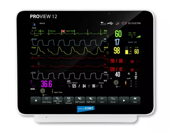
ProView Series ProView 12 patient monitor
ProView Series ProView 12 patient monitor monitor specifications Display: 12.1&Prime...
Portal and digital medical technology fair of the largest MedTech cluster in Germany

ProView Series ProView 12 patient monitor
ProView Series ProView 12 patient monitor monitor specifications Display: 12.1&Prime...
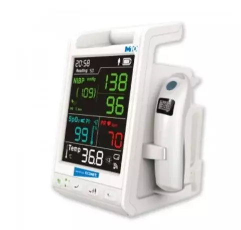
Patient Monitor Vital Signs Monitor M10
Patient Monitor Vital Signs Monitor M10 The compact vital signs monitor M10 was specially developed...
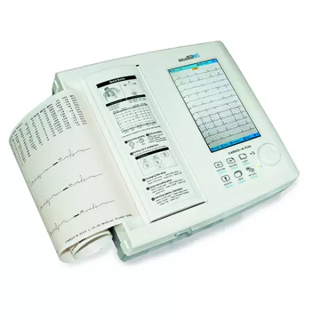
ECG recorder - Cardio-M Plus and Co.
ECG recorder - Cardio-M Plus and Co. 12-lead resting ECG with 7″ touch screen . 7″ touchscreen TFT-L...
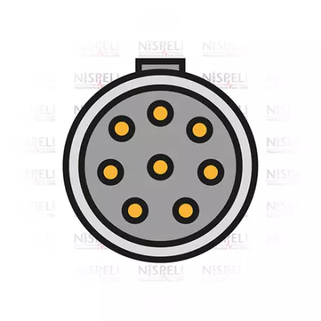
3-wire ECG trunk cable to Philips/HP
3-wire ECG trunk cable to Philips/HP for monitors: 43100A,43110A,43120A,78352C, 78354C,78824C,862474...
3 core complete cable to Philips/HP with...
3 core complete cable to Philips/HP with push button DIN Monitors: C3: 862474, 862475, 862478, 86247...
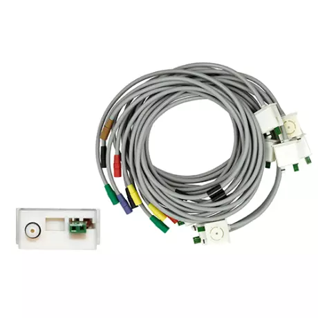
10-core set of electrode lines for the KISS...
10-core set of electrode lines for the KISS suction system Chest wall C1-C6, extremities F, L, R, N
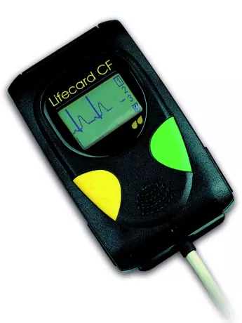
Long-term ECG recorder Lifecard CF
Long-term ECG recorder Lifecard CF Long-term ECG recorder with pacemaker detection and 7-day functio...
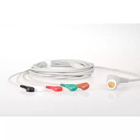
ECG cable systems and connectors
We carry various ECG cable systems and connectors Monitors: C3: 862474, 862475, 862478, 862479, C...
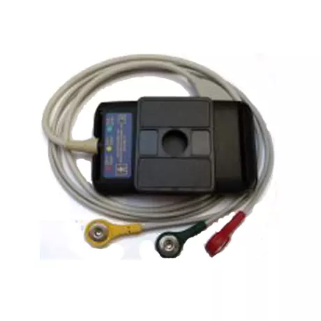
Patient cable for ECG recorder Lifecard CF...
Patient cable for ECG recorder Lifecard CF 3-wire long with clip
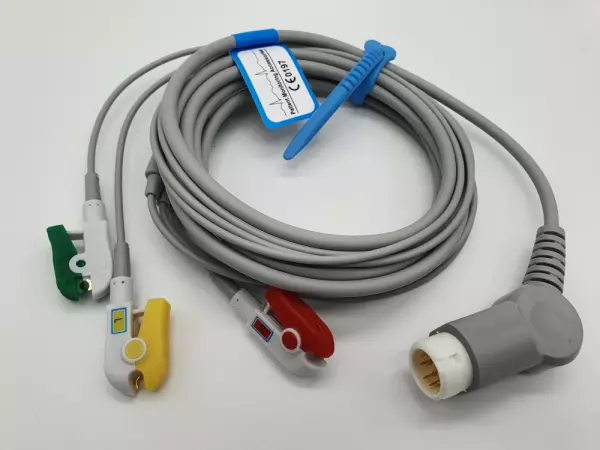
3-wire complete cable to Philips/HP with...
3-wire complete cable to Philips/HP with clamps Monitors: C3: 862474, 862475, 862478, 862479, Codema...
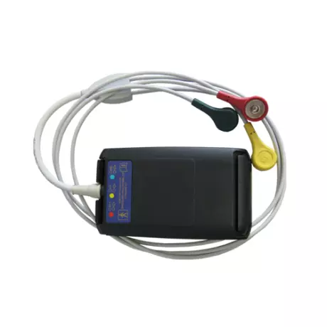
Patient cable for ECG recorder Lifecard CF...
Patient cable for ECG recorder Lifecard CF 3-wire, short
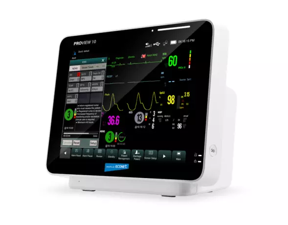
ProView Series ProView 10 patient monitor
ProView Series ProView 10 patient monitor monitor specifications 10.4" TFT full touch display s...
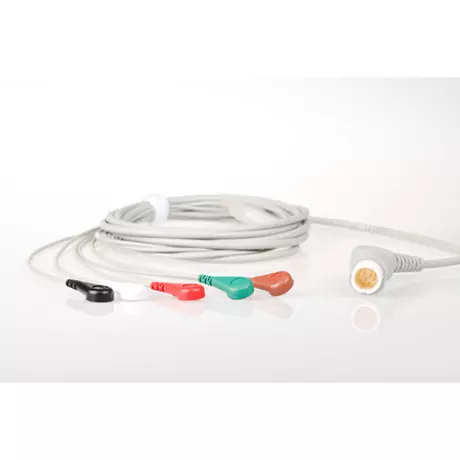
5-lead ECG complete cable to Philips/HP with...
5-lead ECG complete cable to Philips/HP with push button DIN Monitors: C3: 862474, 862475, 862478, 8...

The heart's current is obtained from an electrocardiogram. When the heart beats, the tension in the myocardium has a certain direction and strength. With the help of measuring devices, this voltage can be measured and displayed graphically as a curve (electrocardiogram or EKG for short). If you suspect a heart condition, an electrocardiogram is especially useful. For example, angina pectoris, inflammation of the heart or myocardial infarction show typical changes in the electrocardiogram (EKG). Cardiac arrhythmias can also occur in other diseases, such as B. Hyperthyroidism (hyperthyroidism), which can also appear on the EKG. The electrocardiogram can also be used to check for coronary artery disease (CHD), a previous heart attack if someone has experienced severe pain and chest pressure, or as a routine check-up for routine checkups such as an exam.
But the heart has to keep beating and the brain never tells itself what to do. For this reason it has its own electrical pulse generator: the sinus node. This is where the actual electrical excitation takes place in very special cardiomyocytes. Then the stimulus is transmitted to other myocardial fibers via its own conduction system, so that the heart can contract regularly. Every pumping function of the heart is preceded by an electrical excitation, usually from the sinus node, the main pacemaker of the heart, and the reaching of the muscle cells via the cardiac conduction system. These possible changes in the heart can be recorded by the EKG electrodes on the body surface and recorded on the time axis. The result is a repetitive, relatively even picture of the heart's electrical actions.
In cardiology, in addition to the standard EKG, there are many different EKG procedures that can be used to clarify certain problems, especially when diagnosing arrhythmias. The electrocardiogram does not provide any information about the perfusion (blood flow) of the heart, that is, the condition of the coronary arteries, as long as it does not interfere with the spread of excitation. This is usually only the case with a late blood flow disorder. The classic EKG is performed on a patient who is lying down and relaxed. Hence it is called a resting EKG. This is the opposite of a stress EKG: Here the EKG is recorded on the patient during physical activity on a treadmill or bicycle.
The electrocardiogram provides doctors with information about heart rhythm, frequency, and the production, expansion, and regression of the heart. These diseases usually change with the following diseases: After the EKG exam, the doctor removes the electrodes. The contact gel can be easily removed with paper towels without leaving any residue. In principle, no specific preventive measures were observed. The doctor will use the records to explain your results to you and, if necessary, discuss treatment options with you. For most diagnoses, the ECG only provides information and cannot be evaluated independently of the clinical situation (e.g. myocardial infarction, signs of hypertrophy, myocarditis). Only in the case of arrhythmia or excitation can a clear diagnosis be made on the basis of the electrocardiogram alone.
Become a digital exhibitor yourself in the online portal of the largest and best-known MedTech cluster region in Germany and inform the world of medical technology about your products and services as well as about news, events and career opportunities.
With an attractive online profile, we will help you to present yourself professionally on our portal as well as on Google and on social media.