

Repair service provider for your endoscopy...
endorepair – your partner for the repair of your endoscopy devices and ultrasound probes Ve...
Portal and digital medical technology fair of the largest MedTech cluster in Germany
Repair service provider for your endoscopy...
endorepair – your partner for the repair of your endoscopy devices and ultrasound probes Ve...
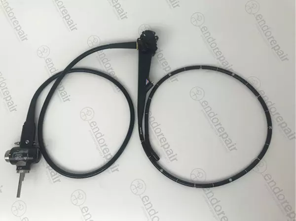
High quality, medical, flexible duodenoscopes...
Our product range of medical endoscopes - High quality, medical, flexible duodenoscopes for exp...
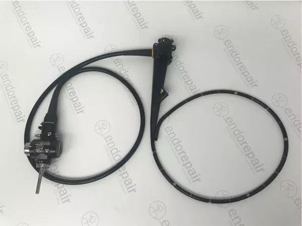
High quality, medical, flexible Gastroscopes...
Our product range of medical endoscopes - High quality, medical, flexible Gastroscopes for expe...
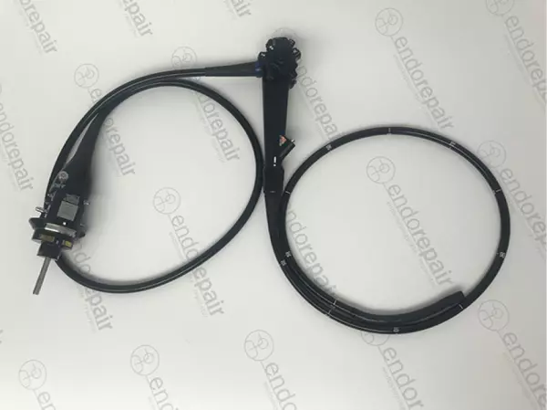
High quality, medical, flexible Coloscopes...
Our product range of medical endoscopes - High quality, medical, flexible Coloscopes for expert...
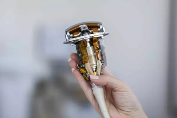
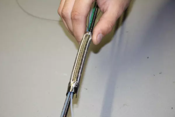
Certified repair services of ultrasonic probes...
endorepair has evolved in the last 2 years and prepared another repair branch. During the two q...
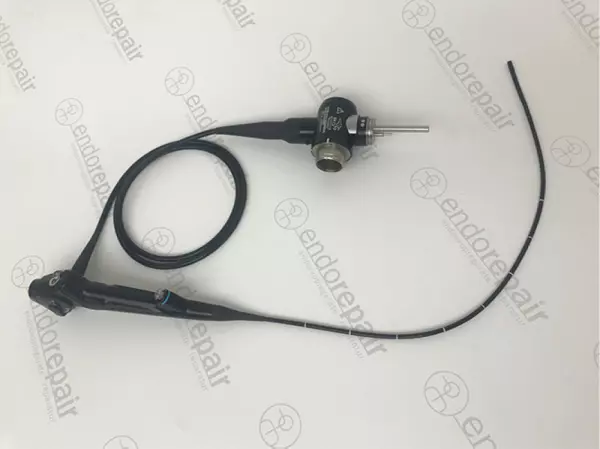
High quality, medical, flexible bronchoscope...
Our product range of medical endoscopes - High quality, medical, flexible bronchoscope for expe...

High quality, medical, flexible ultrasound...
Our product range of medical endoscopes - High quality, medical, flexible ultrasound endoscopes...
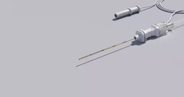

Echogenic, Stimulating Single Shot Nerve Block Needle. SonoPlex needles were especially developed...

Bypassing repairs and rental equipment for...
At endorepair, we help you minimise your economic downtime. We achieve this on the one hand through...


A sonography procedure is a noninvasive method of diagnosing conditions of the body. The technician presses a transducer against the patient's skin to transmit high-frequency sound waves into the body. The transducer measures any subtle changes in these waves. Information resulting from the scan is displayed on a computer. The radiologist interprets the images and performs any necessary treatment. For more information on sonography, contact your doctor today.
This diagnostic imaging technique uses high-frequency sound waves to examine internal body structures. The frequency of the waves varies depending on the size of the internal structures. When they hit these structures, they bounce back as echoes. The sound waves are processed by a computer to produce images called sonograms. The procedure uses a special machine to transmit and receive these sound waves. The images produced are known as sonograms. They allow physicians to view the internal structures of the body.
Ultrasound machines have become commonplace in modern healthcare. They are used to scan a patient's lungs, abdomen, and heart. A sonography technician places an ultrasonic probe on the patient's body. These devices emit ultrasonic waves that can measure the size of the lungs, kidneys, and heart. The transducer is placed near the body to detect the size of an organ. During a sonography procedure, a transducer focuses on a specific area, such as the breast.
During the procedure, a sonographer places a transducer on the area to be imaged. These waves create echoes when they bounce back and are sent to an ultrasound machine. These echoes are converted into images by the machine. Once the images are created, the technician sends them to a computer where they are transformed into pictures. During the procedure, the patient can remain in their regular clothes. There are no side effects or special instructions needed for a sonography procedure.
The process of a sonography is simple. The sonographer applies a colourless gel to the affected area and then positions a transducer over the patient's skin. The transducer then moves over the skin to reflect the ultrasound waves. As the ultrasound waves bounce, they form images on the monitor. The doctor examines the images and makes a diagnosis. If the image is clear and has no irregularities, the patient is healthy.
The pleural line is a ridge that forms a normal lung surface. The parietal pleura is pushed together to form the pleural line. This is the basic principle of a lung ultrasound. The pleural line is located less than a centimeter below the ribs in most adults. It is visible as a horizontal line of hyperechoic light. During a sonography, the pleural ridge is the target area for the examination.
The images produced by a sonography are the same as the images produced by an ultrasound. It is important to understand that the sonogram is not an ultrasound. Rather, it is the image that results from an ultrasound. It is a digital image that is made to show the fetus inside the uterus. However, the image is not the same as a normal baby. The fetal tissue is not present in the ultrasound, and the fetus is not visible.
A single technician performs the test. A patient is required to undress so that the area of interest can be seen. A conductive gel is applied to the skin by the technician. The transducer may be warm or cool. During the test, a transducer may be positioned against the patient's skin to obtain images of the internal organs. When a diagnostic medical sonogram is completed, it will provide physicians with information they need to make the best possible diagnosis.
The use of ultrasound is a great way to diagnose conditions of the body. Besides being an important tool for diagnosing diseases, sonography is also used to monitor the blood flow patterns in the body. This is an imaging technology that makes it possible to visualize organs, including the organs and tissues of the body. It is a noninvasive, safe, and portable method of diagnosis. Moreover, it is a versatile diagnostic tool and can be used in a variety of medical settings.
The purpose of sonography is to monitor the development of a pregnancy. Other uses of the technology include the evaluation of breast lumps, joint and bone conditions, and other medical conditions. An ultrasound may also help guide the needles during biopsies. Regardless of the use of ultrasound technology, it is an essential part of the healthcare field. Hence, it is an indispensable tool for a physician. When a person undergoes an ultrasound, they are able to evaluate the growth of the baby.
Become a digital exhibitor yourself in the online portal of the largest and best-known MedTech cluster region in Germany and inform the world of medical technology about your products and services as well as about news, events and career opportunities.
With an attractive online profile, we will help you to present yourself professionally on our portal as well as on Google and on social media.
